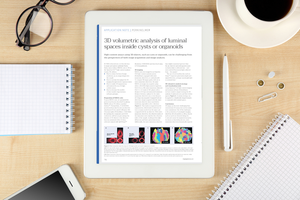
Technical note: 3D volumetric analysis of luminal spaces inside cysts or organoids
Posted: 6 June 2018 | PerkinElmer | No comments yet
High-content assays using 3D objects, such as cysts or organoids, can be challenging from the perspectives of both image acquisition and image analysis.
In this technical note, we describe how to image and analyse epithelial Madin-Darby canine kidney (MDCK) cysts in 3D on the Operetta CLS high-content analysis system.
We address:
- How to reduce the loss of image resolution, as you image deeper into the sample
- How to increase throughput for 3D imaging through shorter exposure times
- How to enable 3D segmentation and analysis for your 3D high-content assays
- How to decrease the number of data transfer steps between software packages.
This technical note is restricted - login or subscribe free to access


Why subscribe? Join our growing community of thousands of industry professionals and gain access to:
- quarterly issues in print and/or digital format
- case studies, whitepapers, webinars and industry-leading content
- breaking news and features
- our extensive online archive of thousands of articles and years of past issues
- ...And it's all free!
Click here to Subscribe today Login here
Related content from this organisation
- Global high-content screening market set to be worth $2.52bn by 2030
- Assays for protein degradation therapeutics to accelerate undruggable proteome discoveries
- Lab automation market set to increase at CAGR of seven percent
- Brochure: Biologics workflow solutions brochure
- Brochure: Viral research solutions
Related topics
3D printing, Assays, Imaging, Screening
Related organisations
PerkinElmer


