Protein offers promise for treatment of reproductive conditions
Posted: 25 October 2023 | Ellen Capon (Drug Target Review) | No comments yet
Scientists found that CXCR4 protein expression outside of the uterus is important for pregnancy maintenance.


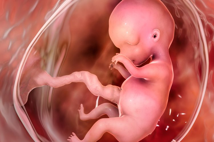
A study led by Yale School of Medicine’s Dr Reshef Tal discovered that a specific protein found throughout the body plays an important role in immune system modulation, influencing placental health early in pregnancy. Their findings may result in new treatments for reproductive conditions.
A human foetus contains genetic material from both parents, which makes it partly foreign to the pregnant person’s body. Therefore, the immune system needs to make adjustments that tolerate the developing foetus whilst protecting against harmful foreign bodies like viruses.
Many of these adaptations happen within the decidua, tissue that surrounds and interacts with the placenta. Previous studies by the same researchers showed that early in pregnancy, bone marrow cells, including immune cells, migrate to the uterus and the decidua. The researchers were intrigued by the mechanisms that recruit these cells to the pregnant uterus.
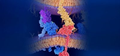
Prior research has shown that a protein called C-X-C chemokine receptor type 4 (CXCR4), and the protein that binds to it are vital for moving bone marrow cells in organs and tissues around the body. CXCR4 is also expressed in higher amounts in the uterus at the beginning of pregnancy.
Dr Tal, Assistant Professor of Obstetrics, Gynaecology and Reproductive Sciences at Yale School of Medicine and senior author of the study, said: “Previous findings imply there is an important function for CXCR4 in pregnancy.” He continued: “We wanted to understand its overall role in pregnancy maintenance and immune function in the decidua.”
To begin, the researchers deleted the gene that codes for CXCR4 in adult female mice, effectively removing the protein from the body. Compared with normal mice, those without CXCR4 lost more foetuses during pregnancy and had smaller litter sizes. However, when CXCR4 was removed only in the uterus, there were no negative pregnancy effects.
“That finding suggested that it’s not the CXCR4 expression within the uterine cells themselves, but rather it’s the CXCR4 expression from outside of the uterus that plays an important role in pregnancy maintenance,” explained Dr Tal.
Since immune cells are recruited to the uterus from outside the organ, the scientists then looked at how CXCR4 deletion affected immune cell populations in the decidua. Early in pregnancy, most immune cells in the decidua are what are known as “natural killer” cells. These white blood cells destroy diseased cells around the body, but in early pregnancy these natural killer cells in the decidua play a key role in the vast tissue and blood vessel remodelling required for the placenta to properly develop and for the foetus to begin receiving nutrients and oxygen from the parent.
Fewer natural killer cells were moved to the uterus in mice without CXCR4, and those that were clustered abnormally. These natural killer cells also expressed a particular enzyme (granzyme B) at lower levels than normal, resulting in an abnormal inflammatory response in the uterus. Also, the mice had irregular blood vessel arrangement in the placenta and decidua, which could affect the exchange of nutrients between parent and foetus.
The researchers transplanted healthy bone marrow from normal mice into mice without CXCR4 to investigate if these changes were caused by immune cell dysfunction. “We found that this rescued much of the effects,” said Dr Tal.
Mice with transplanted bone marrow had fewer pregnancy losses than those without and had normal levels of natural killer cells, cell distribution, expression of enzymes, and blood vessel arrangement in the placenta.
“One of the main takeaways of the study is that you can harness the bone marrow cells’ ability to home to the uterus and affect both the immune status of the decidua and the vascular remodelling of the placenta,” said Dr Tal.
He stated that this could inform new treatments for reproductive conditions like recurrent pregnancy loss and preeclampsia. Preeclampsia, a hypertensive disorder of pregnancy is estimated to complicate 2–8 percent of pregnancies and remains a principal cause of maternal and foetal morbidity and mortality. Preeclampsia may present at any gestation but is more commonly encountered in the third trimester. Multiple risk factors have been documented, including family history, nulliparity, egg donation, diabetes, and obesity.1
“In these conditions, there is thought to be an imbalance in the immune factors and the factors involved in blood vessel formation,” Dr Tal commented. “Our findings could potentially lead to a cell therapy approach to treat those types of conditions.”
This research was published in JCI Insight.
References
1 English FA, Kenny LC, McCarthy FP. Risk factors and effective management of preeclampsia. Dovepress [Internet]. 2015 March 3 [2022 December 15]; 2015(8):7-12. Available from: https://www.dovepress.com/risk-factors-and-effective-management-of-preeclampsia-peer-reviewed-fulltext-article-IBPC
Related topics
Cell Therapy, In Vitro, Protein Expression
Related conditions
Preeclampsia, Recurrent pregnancy loss
Related organisations
Yale School of Medicine
Related people
Dr Reshef Tal (Yale School of Medicine)


