Building better drugs: how 3D models are shaping pre-clinical development
Posted: 25 April 2025 | Viraj Mehta (Engineer - Sai Life Sciences) | No comments yet
Three-dimensional (3D) models are reshaping pre-clinical drug development by providing more accurate insights into drug safety and efficacy. Explore how these advanced in vitro systems help improve predictions and reduce the risk of failure in early-stage drug discovery.
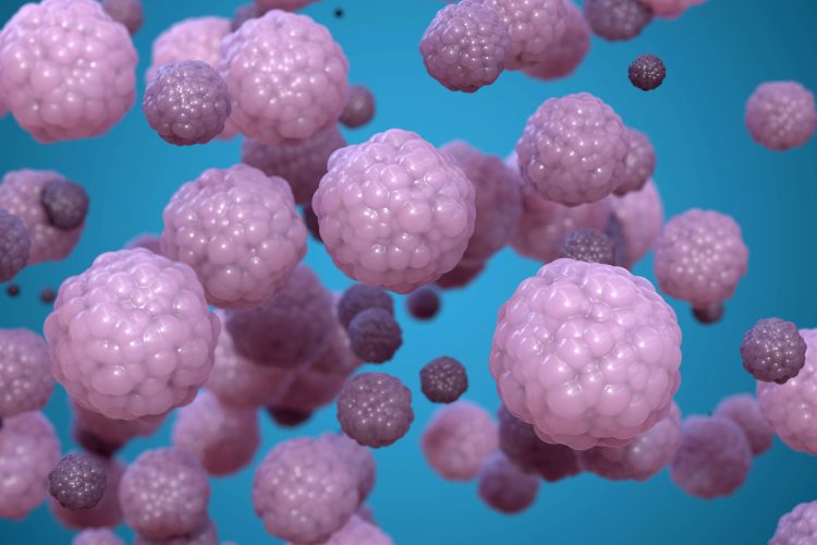
Recent advancements in human-relevant 3D models have paved the way for early predictive insights into pre-clinical drug discovery. The widespread endorsement of new approach methodologies (NAMs) and the principles of the 3Rs (replace, reduce, refine) by regulatory agencies, including the US Food and Drug Administration (FDA) and European Medicines Agency (EMA), has catalysed a paradigm shift in the pre-clinical exploration of biopharmaceutical compounds. Over the past decade, there have been tremendous advancements in the design, micro-manufacturing and biological validation of various complex in vitro models (CIVMs).1 These models, including spheroids, organoids, bioprinted models and organs-on-chips, integrate a combination of 3D microenvironments, primary human-relevant cells, biopolymers derived from tissue matrices, mechanical cues such as stretch and perfusion flow, and micro-patterning.
Prioritised organs and advantages of liver CIVMs
An IQ-MPS survey highlighted that ADME (absorption, distribution, metabolism and excretion) – relevant organs including liver, gut and kidney are the top three prioritised organs of interest across industries to model with CIVMs.2 Liver CIVMs offer several benefits over conventional in vitro systems including prolonged functional activity, enhanced expression of various drug-metabolising enzymes and transporters, and improved cell-cell and cell-ECM (extracellular matrix) interactions.3 These models are pivotal for offering key predictive insights into the clearance of metabolically stable small molecules, maintaining phase I and II metabolic activities for up to four weeks.4
Liver CIVMs offer several benefits over conventional in vitro systems including prolonged functional activity, enhanced expression of various drug-metabolising enzymes and transporters.
Beyond small molecules, liver CIVMs can be leveraged to predict hepatic clearance and identify metabolic soft-spots in molecules that fall beyond rule of five (bRo5) space, including PROteolysis TArgeting Chimeras (PROTACs). PROTACs are a novel class of therapeutic agents that target specific proteins for degradation by harnessing the cell’s natural ubiquitin-proteasome system. Multiple studies have underscored the critical importance of early metabolite identification (MetID) in PROTACs to elucidate the effects of linkers, known to be the most metabolically liable moieties.5,6 Furthermore, the propensity of PROTACs to generate active metabolites that compete with the PROTAC binding site may lead to discrepancies between in vitro and in vivo outcomes.7 Given that the liver is the principal site for PROTAC metabolism, primarily involving CYP3A4 and human aldehyde oxidase (hAOX), human-relevant systems are indispensable for addressing species-specific differences related to aldehyde oxidase.6 With elevated plasma protein binding, the PROTAC modality may also demonstrate significant protein-facilitated uptake. Thus, a system mimicking physiological albumin production rates is essential for accurate hepatic clearance predictions.8
Sai’s matrix-free, high-throughput, multi-well 3D hepatic spheroid-array system can help prospective clients understand the degrader’s hepatic clearance especially when the degrader is metabolically stable. Additionally, the spheroid system can help unravel the propensity for linker cleavage, potential DDI liabilities and potential active metabolites, thus mitigating risks in the early pre-clinical stage of the degrader programme.
Complex in vitro models have evolved over the past decade, using 3D structures, human cells, and mechanical cues to better mimic human tissues.
DILI prediction: comparing spheroids and liver-on-chips
Aside from evaluating the hepatic drug metabolism, liver CIVMs can be used for early predictions of acute and chronic drug-induced liver injury (DILI) liabilities. Emulate’s liver-on-chip with multicellular interactions, dynamic flow and 3D microenvironment have demonstrated the ability to capture species-specific differences in toxicity and intrinsic and idiosyncratic DILI liabilities of small molecules with a sensitivity of 87 percent and specificity of 100 percent.9,10 According to IQ-MPS recommendations, liver-on-chip systems can differentiate between toxic and non-toxic small molecule analogues.10 However, Emulate’s liver-on-chip study reported data from a limited set of compounds with comparatively lower throughput than conventional well-plate-based 3D hepatic models. Addressing the gaps associated with liver-on-chips, a recent 3D hepatic spheroid study predicted DILI liabilities of 152 FDA-approved small molecules using a high-throughput 384 well plate-based system with a sensitivity of 72 percent and specificity of 89 percent.11
Apart from small molecule-associated toxicities, 3D hepatic spheroids can be used to detect DILI liabilities early in antisense oligonucleotides (ASOs).
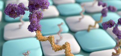
Apart from small molecule-associated toxicities, 3D hepatic spheroids can be used to detect DILI liabilities early in antisense oligonucleotides (ASOs). ASO is a promising drug modality, which primarily prevents mRNA translation into protein through a variety of mechanisms. Hepatic accumulation of ASOs is a significant safety concern, addressable by human-relevant complex in vitro systems. Given their long tissue half-life and human-specific target affinity, long-term 3D human-relevant models are more suitable for detecting ASO-induced toxicities.
FDA-approved ASOs, such as mipomersen and inotersen, received black box warnings due to steatosis detected in late-stage clinical trials. A three-day exposure study conducted by FDA scientists with sandwich-cultured primary human hepatocyte system revealed DILI related to mipomersen, inotersen and their impurities.12 However, the system failed to demonstrate noticeable adenosine triphosphate (ATP) depletion due to the exposure of parent ASOs. Further, hepatocytes in the sandwich model are known to dedifferentiate within two weeks,13 making 3D hepatic spheroids and organoids a better alternative for capturing ASO-associated DILI over repeated drug exposures for one to two weeks. The 3D hepatic spheroid model has accurately captured several N-acetylgalactosamine (GalNAc)-conjugated or locked nucleic acid (LNA)-modified ASO-mediated DILI in two-week studies.14
Apart from ASOs, hepatic safety evaluation of PROTAC-associated ‘bystander degradation’ especially for liver-targeting chimeras (LIVTAC) is also possible using 3D hepatic spheroids. Overall, very few studies have explored CIVMs for safety evaluation of large molecules and biologics, indicating the need for more such validation studies.
Sai’s 3D spheroid model permits repeated drug exposure for up to two weeks to capture potential hepatic toxicities associated with small molecules and new drug modalities, including ASOs. Various readouts are provided, such as cellular ATP-based viability, LDH release, albumin production and ALT production. Additionally, we monitor spheroid morphology and employ high-content imaging-based indicators to elucidate mechanisms associated with DILI, including mitochondrial membrane potential loss, glutathione (GSH) depletion, reactive oxygen species (ROS) accumulation and lipid accumulation. We also provide LC-MS/MS-based reactive metabolite species identification causing DILI.
Expanding CIVMs to kidney and gut models
Aside from hepatotoxicity, FDA-approved ASOs including nusinersen, inoteresen, golodirsen, viltolarsen and casimersen have shown renal toxicity, as they tend to accumulate in renal proximal tubule epithelium. Complex 3D human-relevant kidney models expressing ASO-relevant uptake receptors, such as Megalin and EGFR, can be leveraged to assess their accumulation and downstream toxicity mechanisms.15
In addition to detecting hepatotoxicity and renal toxicity, complex in vitro models in high-throughput 384 well-plate format cultured under perfusion flow can be utilised for detecting drug-induced disruption of the gut epithelium barrier.16 Additionally, such dynamic gut-on-chips offer unprecedented mechanistic insights based on high-content imaging unlike static transwell models.16 These 384 well-plate-based dynamic models can also recapitulate the blood-brain barrier in vitro to understand receptor-mediated transcytosis of large molecules such as therapeutic antibodies.17 Highly human target-specific biologics pose several other safety challenges including antibody drug conjugates (ADCs), adeno-associated virus (AAV) gene therapy and ASO-induced thrombocytopenia, CAR-T-induced cytokine release syndrome, ADC-induced peripheral neuropathies and AAV-induced immune-mediated DILI. Human-relevant complex in vitro models may provide better in vitro-in vivo correlation of such safety risks primarily owing to sustained expression of various human-relevant targets of interest than conventional animal models.18
Highly human target-specific biologics pose several other safety challenges including antibody drug conjugates (ADCs), adeno-associated virus (AAV) gene therapy and ASO-induced thrombocytopenia.
Despite the tremendous potential to detect safety risks of biologics, the lack of regulatory guidelines related to the usage of the complex in vitro models often demotivates industries to report the safety/efficacy data derived fromfassay them. Additionally, proper decision frameworks need to be established to guide industries in implementing CIVMs with animal models to gain confidence and achieve the 3R principles effectively.
Finally, CIVMs address some of the unmet needs in drug discovery, including evaluation of drug transport across the foeto-maternal interface and safety assessment of molecules during pregnancy.19 Multi-organ CIVMs provide several new opportunities in ADME including in vitro bioavailability predictions, assessment of the effect of gut microbiome on hepatic drug metabolism, and prediction of the pharmacokinetic (PK) profile in humans in early pre-clinical stage.3
Despite numerous breakthroughs in validating various organ models, including interconnected multi-organ models, the overall industry adoption rate of human-relevant CIVMs remains low. Various initiatives including FDA Modernization Act 3.0, NIH’s Complement- ARIE programme, NIH’s TraCe MPS grants and the CEN/CENELEC FGOoC Standardization Roadmap have been directed towards standardisation, validation and qualification of CIVMs to increase industry adoption. The recent acceptance of Emulate liver-on-chips into the FDA’s ISTAND programme shows global commitment toward modernising the drug discovery process, reducing drug attrition rates, and a concrete step towards enhancing the adoption of CIVMs in this decade.
References
[1] Strelez C, Jiang HY, Mumenthaler SM. Organs-on-chips: a decade of innovation. Trends Biotechnol 41 (2023) 278–280. https://doi.org/10.1016/j.tibtech.2023.01.004.
[2] Baker TK, Van Vleet TR, Mahalingaiah PK, et al. The Current Status and Use of Microphysiological Systems by the Pharmaceutical Industry: The International Consortium for Innovation and Quality Microphysiological Systems Affiliate Survey and Commentary. Drug Metab Dispos 52 (2024) 198–209. https://doi.org/10.1124/DMD.123.001510.
[3] Mehta V, Karnam G, Madgula V. Liver-on-chips for drug discovery and development. Mater Today Bio 27 (2024) 101143. https://doi.org/10.1016/J.MTBIO.2024.101143.
[4] Preiss LC, Lauschke VM, Georgi K, Petersson C. Multi-Well Array Culture of Primary Human Hepatocyte Spheroids for Clearance Extrapolation of Slowly Metabolized Compounds. AAPS J 24 (2022). https://doi.org/10.1208/S12248-022-00689-Y.
[5] Pike A, Williamson B, Harlfinger S, et al. Optimising proteolysis-targeting chimeras (PROTACs) for oral drug delivery: a drug metabolism and pharmacokinetics perspective. Drug Discov Today 25 (2020) 1793–1800. https://doi.org/10.1016/J.DRUDIS.2020.07.013.
[6] Goracci L, Desantis J, Valeri A, et al. Understanding the Metabolism of Proteolysis Targeting Chimeras (PROTACs): The Next Step toward Pharmaceutical Applications. J Med Chem 63 (2020) 11615–11638. https://doi.org/10.1021/ACS.JMEDCHEM.0C00793
[7] Hayhow TG, Williamson B, Lawson M, et al. Metabolism-driven in vitro/in vivo disconnect of an oral ERɑ VHL-PROTAC. Communications Biology 2024 7:1 7 (2024) 1–17. https://doi.org/10.1038/s42003-024-06238-x.
[8] Da-silva F, Boulenc X, Vermet H, et al. Improving Prediction of Metabolic Clearance Using Quantitative Extrapolation of Results Obtained From Human Hepatic Micropatterned Cocultures Model and by Considering the Impact of Albumin Binding. J Pharm Sci 107 (2018) 1957–1972. https://doi.org/10.1016/j.xphs.2018.03.001.
[9] Jang KJ, Otieno MA, Ronxhi J, et al. Reproducing human and cross-species drug toxicities using a Liver-Chip. Sci Transl Med 11 (2019) eaax5516. https://doi.org/10.1126/scitranslmed.aax5516.
[10] Ewart L, Apostolou A, Briggs SA, et al. Performance assessment and economic analysis of a human Liver-Chip for predictive toxicology. Communications Medicine 2022 2:1 2 (2022) 1–16. https://doi.org/10.1038/s43856-022-00209-1.
[11] Fäs L, Chen M, Tong W, et al. Physiological liver microtissue 384-well microplate system for preclinical hepatotoxicity assessment of therapeutic small molecule drugs. Toxicological Sciences 203 (2024) 79–87. https://doi.org/10.1093/TOXSCI/KFAE123.
[12] Toxicity of three antisense oligonucleotide drugs and eighteen of their impurities in primary human hepatocytes | FDA, (n.d.). https://www.fda.gov/science-research/fda-science-forum/toxicity-three-antisense-oligonucleotide-drugs-and-eighteen-their-impurities-primary-human (accessed March 20, 2025).
[13] Bell CC, Dankers ACA, Lauschke VM, et al. Comparison of Hepatic 2D Sandwich Cultures and 3D Spheroids for Long-term Toxicity Applications: A Multicenter Study. Toxicological Sciences 162 (2018) 655–666. https://doi.org/10.1093/TOXSCI/KFX289.
[14] Hepatotoxicity Assessment of Antisense Oligonucleotides. [Internet]. InSphero. 2024 [cited 2025 Apr 16]. Available from: https://insphero.com/resource/initial-evaluation-of-the-use-of-human-liver-microtissues-for-the-hepatotoxicity-assessment-of-antisense-oligonucleotides/.
[15] Caetano-Pinto P, Haughan K, Kragl A, et al. Epidermal growth factor receptor mediates the basolateral uptake of phosphorothioate-modified antisense oligonucleotides in the kidney, Organs-on-a-Chip 4 (2022) 100022. https://doi.org/10.1016/J.OOC.2022.100022.
[16] Trietsch SJ, Naumovska E, Kurek D, et al. Membrane-free culture and real-time barrier integrity assessment of perfused intestinal epithelium tubes. Nature Communications 2017 8:1 8 (2017) 1–8. https://doi.org/10.1038/s41467-017-00259-3.
[17] Wevers NR, Kasi DG, Gray T, et al. A perfused human blood-brain barrier on-a-chip for high-throughput assessment of barrier function and antibody transport. Fluids Barriers CNS 15 (2018) 1–12. https://doi.org/10.1186/S12987-018-0108-3
[18] Maulana TI, Wevers NR, Kristoforus T, et al. Opportunities for Microphysiological Systems in Toxicity Testing of New Drug Modalities. Annu Rev Pharmacol Toxicol 65 (2025). https://doi.org/10.1146/ANNUREV-PHARMTOX-061724-080621.
[19] Safarzadeh M, Richardson LS, Kammala AK, et al. A multi-organ, feto-maternal interface organ-on-chip, models pregnancy pathology and is a useful preclinical extracellular vesicle drug trial platform. Extracellular Vesicle 3 (2024) 100035. https://doi.org/10.1016/J.VESIC.2024.100035.
 Meet the author
Viraj Mehta is an engineer by training and obtained his PhD in biomedical engineering from the Indian Institute of Technology, Hyderabad (2023). Prior to this he earned his bachelor’s degree in mechanical engineering from NIT-Surat (2018). During his PhD in CIVMs and personalised medicine in cancer, he published six first-author research articles, filed two patents and received several awards, including a national award from the Vice President of India in 2019. He has been a part of Sai Life Sciences since June 2023 and instrumental in establishing CIVMs-based innovative assays in ADME & Toxicology in collaboration with Dr Guruswamy Karnam (Principal Scientist, DMPK) and Dr Vamsi Madgula (Associate Vice-President and HoD, DMPK).
Related topics
Analysis, Analytical Techniques, Assays, Biopharmaceuticals, Cell Cultures, Drug Discovery, Drug Discovery Processes, High-Throughput Screening (HTS), In Vitro, Organ-on-a-Chip, Organoids, Personalised Medicine, Toxicology
Related conditions
Cytokine-release syndrome (CRS), Drug-Induced Liver Injury (DILI), Renal Toxicity, Steatosis
Related organisations
Sai Life Sciences
Related people
Viraj Mehta (Engineer - Sai Life Sciences)