


news
Förster resonance energy transfer development enables imaging of single molecules
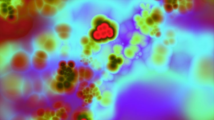
Using fluorescent markers, researchers have developed Förster resonance energy transfer (FRET) to image the assembly, functions and interactions of molecules.
List view / Grid view
Using fluorescent markers, researchers have developed Förster resonance energy transfer (FRET) to image the assembly, functions and interactions of molecules.
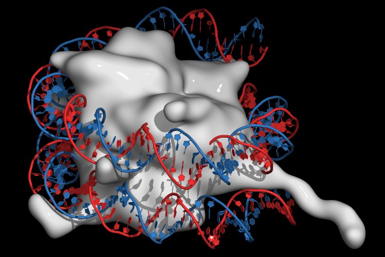
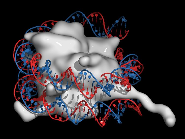
Researchers have shown how ATAD2, a histone chaperone protein, may load histones on to DNA in order to create the chromatin structure.
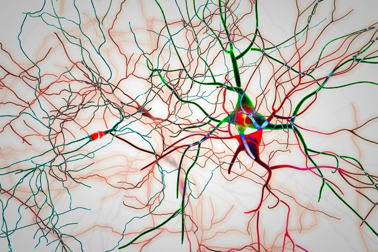
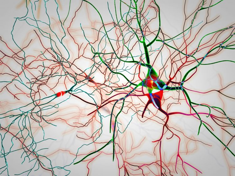
Scientists have identified two master controller regions that are essential for alpha-synuclein aggregation and could be targeted by future therapies.
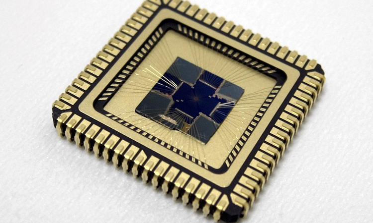
Researchers have applied for a patent for their innovative cantilever and vibrating plate technique which they say could increase the speed of atomic force microscopy on fragile samples.
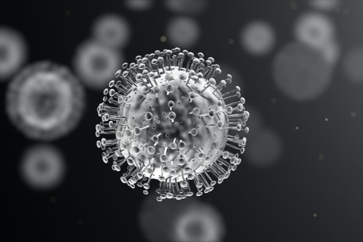
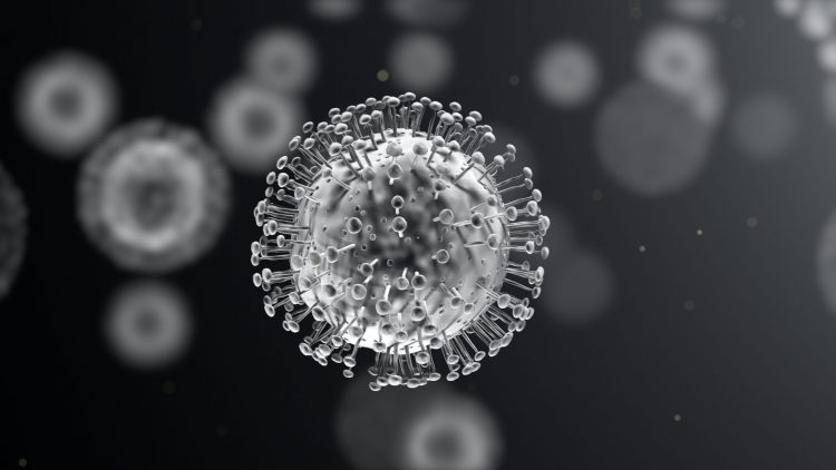
Researchers have used cryogenic electron microscopy to show that coronaviruses enter human cells through an interaction with angiotensin-converting enzyme 2 (ACE2).
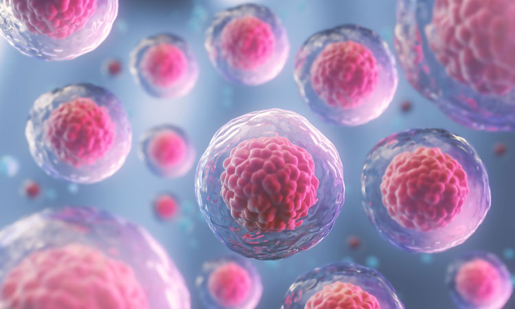
A new imaging technique, which has revealed 3D forces exerted by tiny cell clusters, could help scientists understand how tissue forms, how wounds heal or how tumours spread.
The model was tested on a panel of drugs that are both still on the market or have been recalled due to adverse effects and was able to show their toxicity.
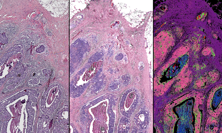
A new method to image cancerous tissues has been created by researchers who have paired infrared measurements with high-resolution optical images.

Using ultrashort laser pulses to interact with vesicles, researchers have created a novel label-free imaging method.
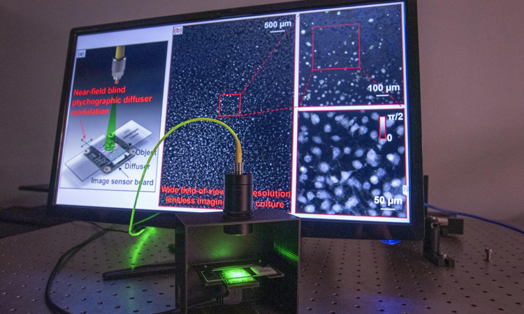
Researchers have developed a new label-free ptychographic microscopy method by bringing samples closer to the image sensor, reducing processing time.
Using cryogenic electron microscopy, a team has mapped the Spike protein on COVID-19, which could be used in the development of vaccines.
Scientists in the US have applied a novel technique to finally unravel a particular kinase enzyme that is associated with familial Parkinson’s disease; providing a clearer potential therapeutic target.
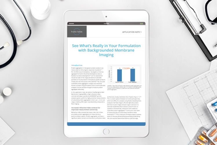
Backgrounded Membrane Imaging (BMI), used by the HORIZON® particle analysis system, performs measurements of subvisible protein aggregates.

A group of researchers, led by Professor Zucai Suo, have revealed the mode of action of two HIV drugs and identified how resistance can develop, which they say could lead to improved drug design in the future.
A label-free imaging technology has been developed by researchers, allowing them to investigate biomolecules such as metabolites, aiding in the study of drugs.