

article
Is an HIV vaccine on the horizon?
Researchers in the US have developed a potential HIV vaccine approach that aims to prompt the creation of broadly neutralising antibodies via mRNA.
List view / Grid view
Researchers in the US have developed a potential HIV vaccine approach that aims to prompt the creation of broadly neutralising antibodies via mRNA.


Join our experts as they discuss the advantages of multiplexed imaging for a wide range of research and how this technique will develop in the future.
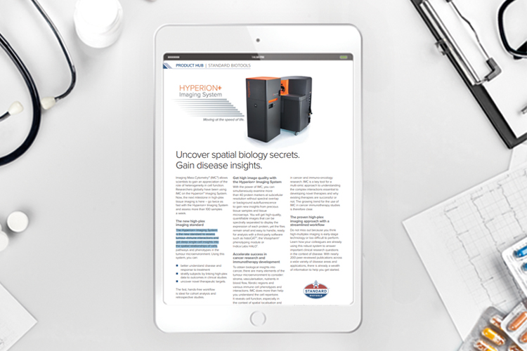
Discover why the Hyperion+ Imaging System is the standard to assess tumour-immune interactions and get deep single-cell insights.

Researchers have successfully characterised a part of the brain that shows the earliest accumulation of tau protein, an important biomarker for the development of Alzheimer's disease.

The researchers created a chronic skull optical clearing window where they no longer needed to remove any piece of the skull.
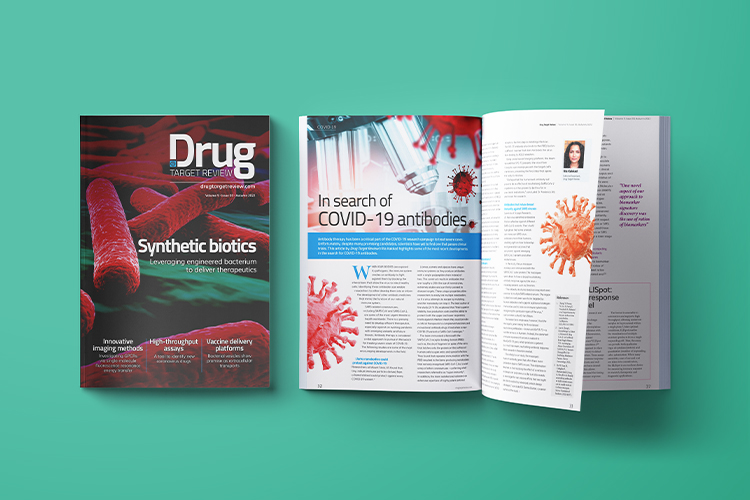
In this issue are articles on synthetically engineered bacteria to deliver therapeutics, how single-molecule fluorescence resonance energy transfer was used to image GPCRs and a new assay to identify coronavirus drugs. Also included are pieces on vaccine development, monoclonal antibodies and neuroscience.
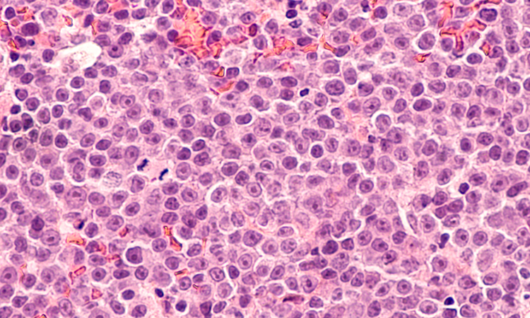
A new high-throughput approach has shown how patients whose tumours express CD58 are more likely to respond to CAR T-cell therapy.

Researchers have developed a novel microscopy technique to make invisible molecules visible by “de-crowding” to expand a cell or tissue sample before labelling the molecules.
Using cryo-electron microscopy, researchers have captured the structure of a membrane-bound T-cell receptor complex with bound antigen.
Researchers have developed a novel label-free method named tomographic phase microscopy in flow cytometry for measuring intracellular lipid droplets in 3D.

R&D Systems offers custom antibody production services to engineer the right antibody to solve your research question with confidence.

Single-domain antibodies (sdAb) are small, stable antibodies derived from camelids with a single monomeric variable domain.
27 July 2022 | By Molecular Devices & HeartBeat.bio
Watch this virtual panel discussion to identify ways to overcome common challenges in 3D biology research, regardless of where you are in your journey.

Researchers for the first time have captured images of an autoantibody bound to a nerve cell surface receptor, revealing the physical mechanism behind a neurological autoimmune disease.
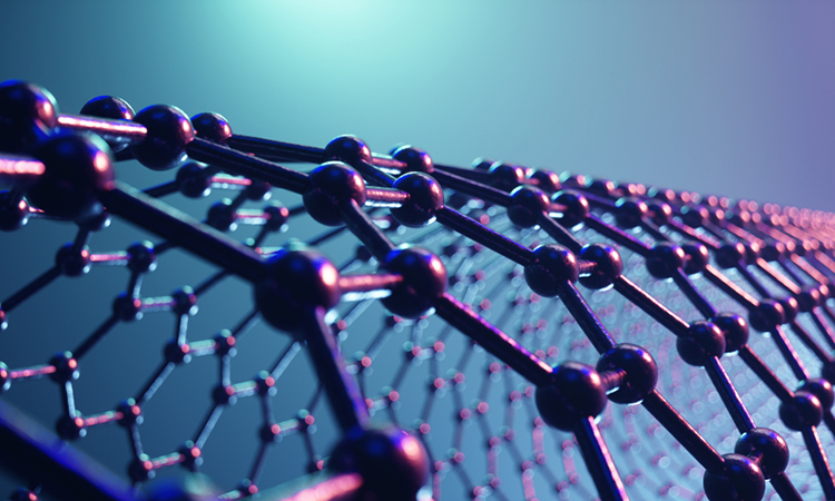
Scientists have created a new imaging technique with graphene which generates clearer pictures of the structures of small molecules.