Novel tool helps differentiate between cancerous and healthy cells for AML
Posted: 25 April 2023 | Izzy Wood (Drug Target Review) | No comments yet
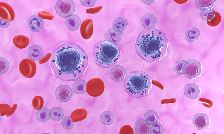
Spanish scientists have developed a new method to identify between cancerous and healthy cells for cases of acute myeloid leukaemia (AML).


Researchers from Centre for Genomic Regulation (CRG), Spain, have made a significant breakthrough in the field of acute myeloid leukaemia (AML) by developing a new method to distinguish between cancerous and healthy stem cells and progenitor cells.
AML is a type of cancer that is driven by malignant blood stem cells and is known for its rapid growth and accumulation of abnormal white blood cells. In the past, identifying these malignant cells has been challenging, leading to difficulties in predicting patient responses to chemotherapy. However, the new findings, published in the journal Cell Stem Cell, offer promising prospects for the development of improved diagnostic and prognostic tools.
The research team utilised a computational method called CloneTracer, which operates at clonal resolution, to analyse data from bone marrow samples of 19 AML patients. The method used a technique known as single-cell RNA sequencing to measure gene expression in thousands of cells simultaneously. CloneTracer revealed two distinct stem cell compartments within the samples: one consisting predominantly of healthy and dormant stem cells, and another highly active compartment mainly composed of leukemic stem cells.
The study also revealed that mutations, including some of the most commonly-mutated AML driver genes, exert their effects primarily in progenitor cells. Progenitor cells are cells that have differentiated to a more mature stage, and their characteristics were found to be correlated with the response of patients to therapy. This finding has important implications for the prognosis and treatment of AML, as progenitor cells that have differentiated to a more mature stage tend to respond better to therapy.
Dr Anne Kathrin Merbach, co-first author of the study and postdoc in the group of Professor Carsten Müller-Tidow at the University Hospital of Heidelberg, explained that by looking at the progenitor cells, they can reasonably predict whether first-line chemotherapy will be successful. However, the researchers acknowledge that one limitation of CloneTracer is that single-cell RNA sequencing is costly and time-consuming, making it unfeasible for routine clinical use. Therefore, they plan to develop alternative ways of characterising the progenitor cells, such as using fluorescence-activated cell sorting (FACS), a commonly used technique in leukaemia research that is more readily available in most haematology medical departments worldwide.
Despite the promising results, the authors of the study caution that targeted therapies must also focus on the actual leukemic stem cell compartment in order to effectively reduce relapse rates and improve long-term survival. The clonal resolution offered by CloneTracer has allowed the research team to characterise the gene expression signature of this critical cell population, which does not respond well to first-line chemotherapy. However, further validation of the findings with larger sample sizes will be necessary before they can be translated into improved therapies.
The development of this new method to distinguish between cancerous and healthy stem cells and progenitor cells in AML represents a significant advancement in the field of leukaemia research. With further validation and refinement, this method has the potential to contribute to the development of personalised treatment strategies for AML patients, allowing for more accurate prediction of therapy responses and ultimately improving.
Related topics
Chemotherapy, Genetic Analysis, Oncology, RNAs, Sequencing, Targets, Therapeutics
Related conditions
acute myeloid leukaemia (AML)
Related organisations
Centre for Genomic Regulation (CRG)
Related people
Dr Anne Kathrin Merbach, Professor Carsten Müller-Tidow


