Investigating the inflammatory myocardial microenvironment
Posted: 13 February 2024 | Drug Target Review | No comments yet
By studying the molecular cell states within transplanted paediatric hearts, researchers have unlocked new treatment strategies.


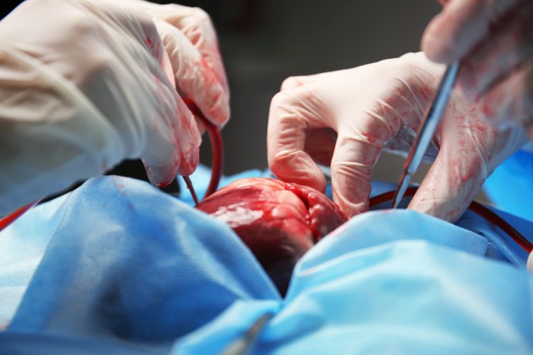
Scientists from The Texas Heart Institute (THI), Baylor College of Medicine, Texas Children’s Hospital and the University of Texas Health Science Center McGovern Medical School have discovered the underlying molecular cell states within transplanted paediatric hearts. This will improve treatment strategies and enhance the longevity of cardiac allografts.
Paediatric heart transplantation is a life-saving intervention for children suffering from end-stage heart failure. However, although the procedure offers hope, the long-term outcomes for patients are suboptimal because of allograft rejection and graft failure.
The research team utilised a dataset comprising of rare heart samples from repeat heart transplantations. Single-nucleus RNA sequencing (snRNA-seq) techniques allowed them to investigate the inflammatory myocardial microenvironment within human paediatric cardiac allografts.
Dr James Martin, physician scientist at THI, Vivian L. Smith Chair in Regenerative Medicine and Vice Chairman and Professor of Integrative Physiology at Baylor College of Medicine, is one of the leaders of the study. He commented: “Our approach offers an unprecedented level of detail…We were able to distinguish immune cells originating from the donor versus the recipient by leveraging naturally occurring genetic variants embedded within our sequencing data. This helps us gain a comprehensive understanding of the immune response dynamics within transplanted hearts.”
This study denotes the first-ever description of molecular cell states within a transplanted paediatric heart at single-cell resolution, examining samples collected as early as five days post-transplantation and extending up to 12 years thereafter. The researchers observed a rapid loss of donor-derived tissue-resident macrophages, which are crucial for graft acceptance and long-term success. Contrastingly, macrophages derived from the recipient’s circulation quickly populated the heart shortly after transplantation. This imbalance between donor-derived and recipient-derived macrophages significantly contributed to allograft failure.
Dr Xiao Li, THI Faculty and Assistant Investigator of the McGill Gene Editing Lab at THI and one of the leaders of the study elucidated: “These findings have significant clinical implications…By targeting the heightened inflammatory response mediated by recipient-derived macrophages and natural killer cells, we can potentially prevent early graft failure and acute rejection episodes. Additionally, preserving the population of resident macrophages within the transplanted heart could pave the way for novel immunomodulation strategies and greatly enhance the longevity of paediatric cardiac allografts.”
Dr Diwakar Turaga, paediatric cardiac intensivist at Texas Children’s Hospital and assistant professor of paediatrics critical care at Baylor College of Medicine, added: “In the CICU, I take care of children who come in with heart rejection. Our medical therapies to treat rejection are still very limited. This study is a major step towards targeted immune therapies and precision medicine.”
These new insights into immune response dynamics could greatly improve patients’ long-term quality and quantity of life.
This study was published in Circulation.
Related topics
Precision Medicine, Regenerative Medicine
Related conditions
allograft rejection, end-stage heart failure, graft failure


