New silk-based membrane for organ-on-a-chip platform
Posted: 7 June 2024 | Drug Target Review | No comments yet
The development of a new membrane which better mimics human extracellular membranes will enable more accurate disease research.
A silk-based, ultrathin membrane that can be used in organ-on-a-chip models have been developed by biomedical engineers at Duke University. These membranes better mimic the natural environment of cells and tissues within the body, which will enable researchers to more accurately replicate a wide range of diseases and test therapeutics.
Organ-on-a-chip (OOC) systems have been revolutionary, in terms of producing dynamic models of tissue structures, investigating organ functions or modelling diseases. Scientists can populate these tools with human stem cells to generate patient-specific organ models for preclinical studies. However, challenges in the chip’s design have emerged, particularly with the materials used to make the membranes that form the support structure for specialised cells to grow on.
Normally, membranes are composed of polymers that do not degrade, creating a permanent barrier between cells and tissues which is between 30 to 50 microns thick, impacting communication between cells and limiting cell growth. Comparatively, extracellular membranes in human organs are often less than one micron thick.
Dr Samira Musah, Assistant professor of biomedical engineering and medicine at Duke, explained: “We want to handle the tissues in these chips just like a pathologist would handle biopsy samples or even living tissues from a patient, but this wasn’t possible with the standard polymer membranes because the extra thickness prevented the cells from forming structures that more closely resemble tissues in the human body…We thought, ‘Wouldn’t it be nice if we could get a protein-based material that mimics the structure of these natural membranes and is thin enough for us to slice and study?’”
This prompted Dr Musah and George (Xingrui) Mou, a PhD student in Dr Musah’s lab and first author on the paper, to investigate silk fibroin, a protein made by silkworms that can be electronically spun into a membrane. Having previously been used to form scaffolds for wound healing, its long, intertwining fibres and porosity better mimics the structure of the extracellular matrix seen in human organs.
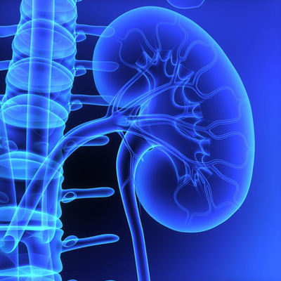
Kidney disease
The new membrane, which was five or fewer microns, was applied to kidney chip models. This OOC platform is meant to resemble a cross section of the glomerular capillary wall, an essential structure in kidneys formed from clusters of blood vessels, which is responsible for filtering blood.
Human induced pluripotent stem cells (hiPSCs) were added into the chip once the membrane was in place. The team observed that these cells could send signals across the ultrathin membrane, helping the cells to differentiate into glomerular cells, podocytes and vascular endothelial cells. Furthermore, the platform initiated the development of endothelial fenestrations in the growing tissue, holes that allow for the passage of fluid between the cellular layers. The different kidney cell types had assembled into a glomerular capillary wall by the end of the test and could efficiently filter molecules by size.
Due to there being a lack of effective models for kidney disease, despite it affecting over 15 percent of American adults, Dr Musah and his colleagues are aiming to use this technology to elucidate the mechanisms driving it. “Using this platform to develop kidney disease models could help us discover new biomarkers of the disease,” commented Mao. “This could also be used to help us screen for drug candidates for several kidney disease models.”
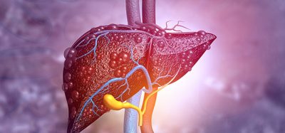
“This technology has implications for all organ-on-a-chip models…Our tissues are made up of membranes and interfaces, so you can imagine using this membrane to improve models of other organs, like the brain, liver, and lungs, or other disease states. That’s where the power of our platform really lies.”
This study was published in Science Advances.
Related topics
Disease Research, Organ-on-a-Chip, Stem Cells, Therapeutics
Related conditions
Kidney disease
Related organisations
Duke University