New 3D genomic profiling technique details PanINs
Posted: 19 June 2024 | Drug Target Review | No comments yet
Researchers have developed a 3D approach to improve the characterisation of pancreatic intraepithelial neoplasias.
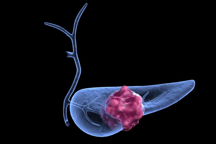
A three-dimensional (3D) genomic profiling technique to identify pancreatic intraepithelial neoplasia’s (PanINs), that result in one of the most aggressive pancreatic cancers, has been developed by scientists at the Johns Hopkins Kimmel Cancer Center’s Sol Goldman Pancreatic Cancer Research Center. The novel technique offers the most detailed 3D map of precancerous lesions in the human pancreas to date.
Dr Laura Wood, one of the study’s senior authors and associate professor of pathology and oncology at the Kimmel Cancer Center and the Johns Hopkins University School of Medicine, commented: “Not many people actually develop pancreatic cancer, so we were shocked to find a lot of precancer, or PanINs, within the normal regions of the pancreas…This research highlights what we don’t yet know about normal aging and raises fundamental questions about how cancer arises in the human pancreas.”
PanINs are difficult to detect and cannot be found by a normal radiology examination due to their small size. This frequently means that when patients receive a diagnosis, the cancer is already advanced and has metastasised to other organs. Current two-dimensional (2D) histological staining methods only provide a limited view of PanINs, so to improve their characterisation, the team sought to create a 3D approach.
“One of the ways we can make a difference for the largest number of people with cancer is through prevention”
CODA, a machine learning (ML) pipeline was developed by analysing 38 normal pancreatic samples, which were thinly sliced and stained onto hundreds of sequential 2D slides. The slide images were reconstructed into digital 3D images, which showed complex networks of interconnected PanINs. The PanINs had an average overall burden of 13 per cubic centimetre, and a range of from one to 31 per cubic centimetre. Also, individuals with pancreatic ductal adenocarcinoma (PDAC) in other regions of their pancreas seemed to have a higher PanIN burden than those with nonductal disease; however, this was not statistically significant.
Furthermore, the team investigated eight of the samples by 3D-guided microdissection and DNA sequencing of specific PanINs. Genomic analysis showed that the networks consisted of genetically distinct PanINs driven by different gene mutations, like mutations in the cancer-causing gene Kirsten rat sarcoma virus (KRAS), found in most pancreatic cancers. Dr Wood noted that the result of multiple precancerous lesions arising from independent mutations is something that has not yet been seen in other organs. However, she said that this discovery will enable scientists to target PanINs, like through KRAS.
An advantage of CODA, as assistant professor of pathology at Johns Hopkins University School of Medicine Dr Ashley Kiemen stated, is that it can be applied to any tissue, disease or model organism, despite it not being usable in a diagnostic setting presently. She added: “We want to continue to investigate what this means in the context of other organ tissues. If normal tissue has thousands of PanINs, then how do we identify which ones are clinically relevant to disease and which ones are not?”
Dr Wood concluded: “One of the ways we can make a difference for the largest number of people with cancer is through prevention, and better understanding the early precursors to cancer through detailed and anatomic molecular maps is the first step,”
This study was published in Nature.
Related topics
Cancer research, Drug Targets, Genomics, Machine learning, Oncology, Radiotherapy
Related conditions
Cancer Research, Pancreatic ductal adenocarcinoma (PDAC)
Related organisations
Johns Hopkins Kimmel Cancer Center, Sol Goldman Pancreatic Cancer Research Center