The real-time capture of transcription complex formation
Posted: 8 July 2024 | Drug Target Review | No comments yet
Researchers have obtained images with a previously unseen level of detail, demonstrating how RNA polymerase opens the transcription bubble.
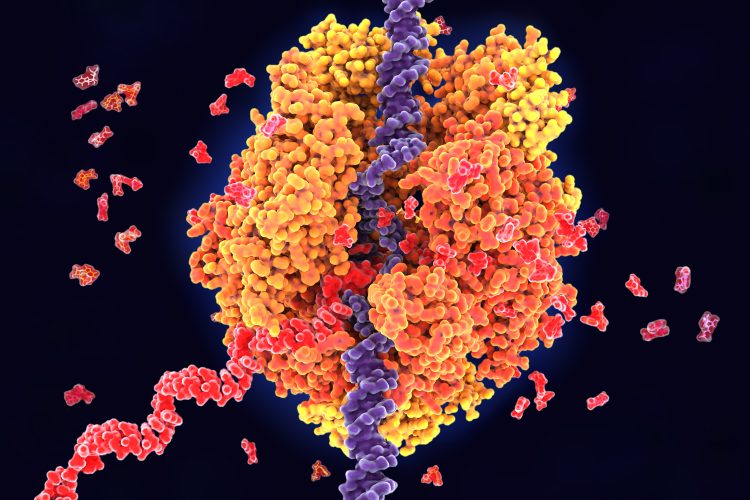
Researchers at The Rockefeller University have captured, for the first time, how RNA polymerase (RNAP) opens the transcription bubble. Within 500 milliseconds of RNAP mixing with DNA, the fundamental workings of transcription were shown, which answers pervading questions about the initiation mechanism and the importance of its various steps.
DNA is transcribed into RNA in every living cell. The transcription process starts when RNAP locks onto DNA. In a few hundred milliseconds, the DNA double helix unwinds to form a node called the transcription bubble, for the exposed DNA strand to be copied into a complementary RNA strand.
Dr Ruth Saecker, first author and research specialist in Dr Seth Darst’s laboratory at Rockefeller, stated: “This is the first time anybody has been able to capture transient transcription complexes as they form in real time…Understanding this process is crucial, as it is a major regulatory step in gene expression.”
RNAP
Dr Darst was the first to describe the structure of bacterial RNAP, and detailing this has been a key focus of his lab. Although years of work have determined that RNAP binding to a specific sequence of DNA initiates a series of steps that open the bubble, the question of how RNAP separates the strands and positions one strand in its active site prevailed.
Dr Andreas Mueller, co-author and postdoctoral fellow in the lab, explained: “We knew from other biological techniques that, when RNAP first encounters DNA, it makes a bunch of intermediate complexes that are highly regulated…But this part of the process can happen in less than a second, and we were unable to capture structures on such a short timescale.”
Cryo-electron microscopy analysis
Collaborating with a team at the New York Structural Biology Center, who developed a robotic, ink-jet system that could quickly prepare biological samples for cryo-electron microscopy analysis, they observed complexes forming in the first 100 to 500 milliseconds of RNAP meeting DNA. This provided images of four distinct intermediate complexes in enough detail to enable analysis. Dr Saecker commented: “The technology was extremely important to this experiment…Without the ability to mix DNA and RNAP quickly and capture an image of it in real-time, these results don’t exist.”
By studying the images, the researchers outlined a sequence of events demonstrating how RNAP interacts with the DNA strands as they separate. As the DNA unwinds, RNAP gradually holds one of the DNA strands to stop the double helix from rejoining. Each new interaction causes RNAP to change shape, allowing more protein-DNA connections to form, including pushing out one part of a protein that prevents DNA from entering RNAP’s active site and forming a stable transcription bubble.
Moving forwards, the team hopes to investigate different stages in transcription and confirm a new hypothesis: that the rate-limiting step in transcription may be the positioning of the DNA template strand within the active site of the RNAP enzyme.
The study’s findings show the value of being able to capture molecular events happening within milliseconds in real-time. “If we want to understand one of the most fundamental processes in life, something that all cells do, we need to understand how its progress and speed are regulated,” concluded Dr Darst. “Once we know that, we’ll have a much clearer picture of how transcription begins.”
This study was published in Nature Structural & Molecular Biology.
Related topics
DNA, Enzymes, Microscopy, Molecular Biology, RNAs
Related organisations
New York Structural Biology Center, The Rockefeller University