New model offers a unique method to study Parkinson’s disease
Posted: 24 July 2024 | Drug Target Review | No comments yet
Mice with rod-specific VPS35 deletion demonstrate a pathology more similar to human Parkinson’s disease, compared to other mouse models.


Researchers at Weill Cornell Medicine have developed a new preclinical model which offers a unique method to study the process of, and detect, Parkinson’s disease (PD). The team demonstrated that removing a crucial component involved in protein transportation in the light-sensing rod cells of mice results in the retinal accumulation of the aggregates of the alpha-synuclein protein found in patients with PD.
Dr Ching-Hea Sung, the Betty Neuwirth Lee and Chilly Professor in Stem Cell Research and a professor of cell biology in ophthalmology and of cell and developmental biology at Weill Cornell Medicine, elucidated: “This is a really unique model involving a pathology that seems more like human Parkinson’s than what we see in other mouse models.”
PD is the second most common neurodegenerative disease after Alzheimer’s disease (AD), affecting around one million Americans. Despite it being primarily known as a movement disorder, its effects on the brain and body are widespread and can include early vision problems and sleep disorders.
Engineered mice
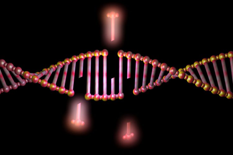
In the study, the scientists engineered mice that lack the gene for the VPS35 protein in rod cells. VPS35 is known for helping cells to distribute molecules, including sending abnormal proteins for degradation. A familial form of PD has been linked to a mutation in VPS35’s gene.
It was observed, even in young mice, that the rods lacking VPS35 quickly lost their synapses, which led to visual impairment similar to that seen in patients with PD. Alpha-synuclein aggregates started to form, and as the affected rods began to die, the mouse retinas showed large, insoluble inclusions that looked like Lewy bodies. Lewy bodies contain alpha-synuclein aggregates and are one of the classic pathological signs of PD.
Dr Sung stated that the results suggest the new model could be very useful for studying disease mechanisms and testing potential therapies. Its advantages include a rapidly developing disease process, as well as the absence of any artificial modification to the mice’s alpha-synuclein. This is unlike existing models that drive pathology using excess, mutant or non-mouse forms of the protein.
Moreover, these results indicate a possible novel strategy for detecting PD. The researchers could use a fundoscope to observe bright areas of autofluorescence caused by lipofuscin, which associate with alpha-synuclein aggregates, even in three-month old mice lacking rod-cell VPS35. Presently, Dr Sung and her international physician collaborators are planning a clinical trial of this approach.
This study was published in in Nature Communications.
Related topics
Animal Models, Drug Discovery Processes, Genetic Analysis, Genome Editing, Neurosciences
Related conditions
Alzheimer's disease (AD), Parkinson's disease (PD)
Related organisations
Weill Cornell Medicine
Related people
Dr Ching-Hea Sung (Weill Cornell Medicine)


