New medicinal advances could be yielded by studying proteins in glass
Posted: 3 July 2019 | Drug Target Review | No comments yet
A unique method for studying proteins has been developed which could open new doors for medicinal research.

The Chalmers researchers have already patented the method. The patent process was unusually fast, indicating the unique character of the method. "Now, with the help of the patent, we can allow the medical industry to use the method. If we didn't have the patent, we would have been less willing to put resources into it because we know now that we can protect forthcoming results," says Martin Andersson (credit: Johan Bodell/Chalmers University of Technology).
A research group at Chalmers University of Technology, under the leadership of Martin Andersson at the Department of Chemistry and Chemical Engineering, has now successfully used Atom Probe Tomography to image and study proteins.
“It was in connection with a study of contact surfaces between the skeleton and implants when we discovered we could distinguish organic materials in the bone with this technique. That gave us the idea to develop the method further,” said Martin Andersson.
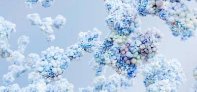
The challenge lay in developing a method of keeping the proteins intact in their natural environment, which the researchers successfully achieved by encapsulating the protein in a thin piece of glass, only around 50 nanometres in diameter (a nanometre is 1/millionth of a millimetre.) They then sliced off the outermost layer of the glass using an electrical field, freeing the protein atom by atom. The protein could then be recreated in 3D on a computer.
Automation now plays a central role in discovery. From self-driving laboratories to real-time bioprocessing
This report explores how data-driven systems improve reproducibility, speed decisions and make scale achievable across research and development.
Inside the report:
- Advance discovery through miniaturised, high-throughput and animal-free systems
- Integrate AI, robotics and analytics to speed decision-making
- Streamline cell therapy and bioprocess QC for scale and compliance
- And more!
This report unlocks perspectives that show how automation is changing the scale and quality of discovery. The result is faster insight, stronger data and better science – access your free copy today
As well as modelling the three-dimensional structure, it simultaneously reveals the proteins’ chemical composition.
“Our method offers a lot of good solutions and can be a strong complement to existing methods. It will be possible to study how proteins are built at an atomic level,” continued Andersson.
With this method, potentially all proteins can be studied: “With this method, we can study individual proteins, as opposed to current methods which study a large number of proteins and then create an average value,” added Gustav Sundell, a researcher in Martin Andersson’s research group.
With Atom Probe Tomography, information on an atom’s mass can also be derived: “Because we collect information on atoms’ masses in our method, it means we can measure the weight,” explained Mats Hulander, a researcher in Martin Andersson’s group. “We can then, for example, create tests where medicinal molecules are combined with different isotopes – giving them different masses – which makes them distinguishable in a study. It should contribute to speeding up processes for constructing and testing new medicines.”
In the future, the researchers will refine the method to improve the speed and accuracy.
The results of the study have been verified through comparison with existing three-dimensional models of known proteins and have published in the scientific journal, Small.
Related topics
Analysis, Protein, Research & Development, Technology
Related organisations
Chalmers University of Technology
Related people
Gustav Sundell, Martin Andersson, Mats Hulander