Technique to visualise hundreds of genomes simultaneously developed
Posted: 28 July 2020 | Victoria Rees (Drug Target Review) | No comments yet
A new technology named OligoFISSEQ has been created which can image and three-dimensionally map the genomes in hundreds of cells at the same time.


Researchers have developed the first technology able to visualise hundreds to potentially thousands of genomes at the same time under a microscope. According to the team, the method images genomes more cheaply, more quickly and increases range of visibility compared to currently available methods.
The study was conducted by researchers at Harvard University, US, the Centro Nacional de Análisis Genómico (CNAG), Spain, and the Centre for Genomic Regulation (CRG), Spain.
Each human cell has two metres of genome condensed down into 10 microns within the cell nucleus. This folds to help genes make physical contact with other genes that may be located a distance away along the chromosome. The researchers say this three-dimensional (3D) organisation is crucial for cell function, but its complexity and constant dynamism make it difficult to visualise. Therefore, imaging more than a handful of genes at the same time has been previously impossible, limiting scientists’ ability to characterise how genomes function.
Reduce preclinical failures with smarter off-target profiling
24 September 2025 | 15:00PM BST | FREE Webinar
Join this webinar to hear from Dr Emilie Desfosses as she shares insights into how in vitro and in silico methods can support more informed, human-relevant safety decisions -especially as ethical and regulatory changes continue to reshape preclinical research.
What you’ll learn:
- Approaches for prioritizing follow-up studies and refining risk mitigation strategies
- How to interpret hit profiles from binding and functional assays
- Strategies for identifying organ systems at risk based on target activity modulation
- How to use visualization tools to assess safety margins and compare compound profiles
Register Now – It’s Free!
However, the researchers developed their technology, named OligoFISSEQ, using new computational methods that overcome the limitations of fluorescence in situ hybridisation (FISH), which can only image a handful of genes at the same time. They used it to create 3D maps of 66 genomic targets across six chromosomes in hundreds of cells, showcasing the method’s potential to visualise the entire genome at a molecular resolution.


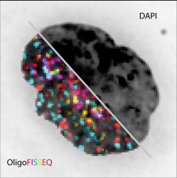
Pictured is the same cell nucleus visualised with conventional fluorescence image (DAPI) compared to with OligoFISSEQ for a 129 different loci [credit: Marc Marti-Renom].
“Up to this moment, seeing a large number of different genes at the same time under the microscope was impossible,” said co-lead author Professor Marc Marti-Renom at CNAG-CRG. “We combined existing sequencing technologies in a smart way so that we can see hundreds of genes by sequencing their targets under the microscope. Before OligoFISSEQ, reaching this number of genes at the same time was slow and expensive. It is like upgrading from a dial-up phoneline to fibre-optic internet and paying 40 times less for it.”
The researchers also tested OligoFISSEQ by mapping all of the 46 regions along the length of the human X chromosome. The higher resolution of the technology revealed new patterns in the way the genome organises itself, including that the length of the chromosome’s arm is not correlated with its angle. The researchers hypothesise this may be indicative of cell type, cell state or cellular health or age.
“The resolution offered by OligoFISSEQ has huge potential to shed new light on patterns we could not see before,” concluded Marti-Renom. “Because of the coverage it provides to study the genome, it is well suited for spotting what may seem like a minor, seemingly inconsequential change in one part of the nucleus that may have a ‘butterfly effect’ elsewhere. What may have previously have been thought of as random may turn out to be anything but. That is the power of this tool.”
The technique is described in Nature Methods.
Related topics
DNA, Genetic Analysis, Genomics, Imaging, Informatics, Targets
Related organisations
Centre for Genomic Regulation (CRG), Centro Nacional de Análisis Genómico (CNAG), Harvard University
Related people
Professor Marc Marti-Renom


