Assay that mimics SARS-CoV-2 infection could test potential COVID-19 drugs
Posted: 22 September 2020 | Victoria Rees (Drug Target Review) | No comments yet
Scientists have developed a new assay that uses nanoparticles to mimic how SARS-CoV-2 infects a cell, which could be used to screen for drugs that treat COVID-19.


A team of scientists has developed a new tool that they say mimics how SARS-CoV-2 – the virus that causes COVID-19 – infects a cell, information that could potentially speed the search for treatments against the disease.
The group are from the National Center for Advancing Translational Sciences (NCATS) and Naval Research Laboratory (NRL), both in the US.
The researchers explain that their tool is a fluorescent nanoparticle probe that uses the Spike (S) protein on the surface of SARS-CoV-2 to bind to human cells and trigger the process that allows the virus to enter the cells. They say the probe could be used in tests to rapidly gauge the ability of biologics, drugs and compounds to block the actual virus from infecting human cells.
“Our goal is to create a screening system to find compounds that block SARS-CoV-2 from binding to cells and infecting them,” explained co-corresponding author of the study Dr Kirill Gorshkov.
The team say that using the actual virus in such screening studies would be difficult and require special facilities. Instead, Gorshkov and Dr Eunkeu Oh, also co-corresponding author of the study and their colleagues wanted to use nanoparticles to mimic the viral function of binding to and invading the host human cell.
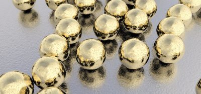
To create the probe, the scientists built a fluorescent nanoparticle called a quantum dot, fashioned from cadmium and selenium. At around 10 nanometres in size, the researchers say these spherical nanoparticles are 3,000 times smaller than the width of a human hair.
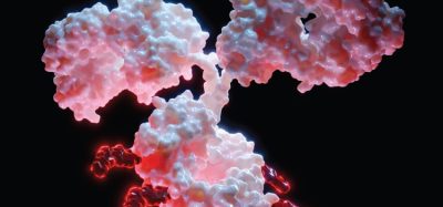
The team then studded the quantum dots’ surfaces with a section of the SARS-CoV-2 S protein that binds to the angiotensin-converting enzyme 2 (ACE2) receptor on human cells. The union of the S protein with ACE2 is the first step in the pathway to viral infection.
The glow from the quantum dots allowed scientists to track the dots’ behaviour under a microscope. “Because they’re such bright fluorescent objects, the quantum dots give us a powerful system to track viral attachment and effects on the cell in real time,” explained Gorshkov.
The investigators tracked how the quantum dot probes interacted with human cells that have ACE2 on their surfaces. They watched the nanoparticle probes attach to ACE2, which combined with the probes and pulled them into the cells. The quantum dot probes did the same in a lung cell line commonly used in coronavirus assays. Safety data showed that the probes were not toxic to the test cells at the concentrations and exposure times used in the study.
The quantum dots followed the SARS-CoV-2 pathway into cells, but the researchers found the probes also mimicked the virus in the presence of antibodies. The antibodies proved to be potent inhibitors of the quantum dot probes, preventing them from binding to ACE2 and entering human cells.
That antibody response means the quantum dot probes could help researchers rapidly test the ability of potential therapeutic agents to block the virus from entering and infecting cells. Assays using the probes could also determine the concentrations at which potential treatments may safely and effectively block infection.
“Using the quantum dots, we could create tests to use in drug screening and drug repurposing, using libraries of compounds that have activity but that also are approved by the US Food and Drug Administration (FDA),” Gorshkov said. “Such assays could rapidly identify promising, safe treatments for COVID-19.”
The researchers highlight that ACE2 may not be the only receptor SARS-CoV-2 targets so the quantum dot probe’s flexible design will allow researchers to swap in proteins that bind to other receptors. With the probe, researchers could also test how mutations in the S protein change the way the virus behaves and how well treatments work by adding the mutated S to the quantum dots.
The study was published in ACS Nano.
Related topics
Assays, Drug Discovery, Drug Discovery Processes, Drug Targets, Nanoparticles, Nanotechnology, Research & Development, Screening
Related conditions
Covid-19
Related organisations
National Center for Advancing Translational Sciences' (NCATS), Naval Research Laboratory (NRL)
Related people
Dr Eunkeu Oh, Dr Kirill Gorshkov


