Scientists reveal ultrastructure of Huntington’s disease
Posted: 15 November 2021 | Anna Begley (Drug Target Review) | No comments yet
Researchers used advanced microscopy and proteomics to unveil new findings on Huntington’s disease and open avenues to future treatments.


A new study from the Ecole Polytechnique Fédérale de Lausanne (EPFL), France, have carried out the first study that interrogates both the ultrastructure of huntingtin inclusion bodies and their composition in cells that model the pathological formation of Huntington’s disease.
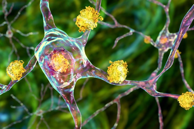
Huntington’s is caused by a mutation in the gene that encodes huntingtin, a protein that normally plays important roles in keeping brain cells healthy and active. However, the mutation “gives” huntingtin an abnormally long tail of glutamine amino acids which cause huntingtin to aggregate inside neurons and eventually kill them. These aggregates have been the subject of a lot of research efforts in the attempt to find a way to understand and treat Huntington’s, but there has been a lack of deep analyses into the inclusions’ ultrastructure.
NEWS: DNA mechanism discovery may open up Huntington’s disease therapies
READ HERE
Using advanced microscopy and proteomics, scientists led by Professor Hilal Lashuel at EPFL have now uncovered this ultrastructure. The researchers used an advanced technique known as correlative light-electron microscopy (CLEM) that can identify inclusions in different cell compartments and study their structural and organisational features of inside cultured neurons. CLEM essentially combines an electron microscope with a fluorescence microscope.
The researchers also used electron tomography which allowed them to visualise individual aggregates within these inclusions. “We wanted to go beyond just painting a detailed picture of how these inclusions form,” explained first author Dr Nathan Riguet. “This is why we extended our studies to understand how these inclusions form, how they impact their subcellular environment, the function of organelles in their vicinity, and ultimately the viability of neurons.”
The study, published in Nature Communications, found that the aggregation of huntingtin and the formation of inclusions in the cytoplasm and nucleus of neurons occurs through different mechanisms, lead to distinct biochemical and ultrastructural properties. The team stated that this means these two types of inclusions may also be toxic through different ways so treatments must employ different strategies to address their formation, maturation, and toxicity.
According to the researchers, the study also suggests that finding ways to modify the growth of inclusions and their interactions with other proteins and organelles is an alternative or complementary strategy for slowing the progression of Huntington’s, especially after disease onset as this process leads to the sequestration of functional proteins and disrupts the function of important organelles in the cell such as the mitochondria.
“Our findings also caution against the use of large fluorescent protein tags to develop disease models of Huntington’s disease and screen for drugs that modify Huntingtin aggregation and inclusion formation,” added Lashuel. “This has significant implications for Huntington’s disease and other neurodegenerative diseases, where fusion of fluorescent proteins is commonly used to investigate mechanisms of protein aggregation and in drug discovery.
Lashuel believes that the study opens up new insights into the composition and ultrastructure of huntingtin inclusions and point to new directions for therapeutic interventions which the team plan to pursue.
Related topics
Microscopy, Molecular Biology, Neurons, Protein, Protein Expression, Small Molecules
Related conditions
Huntington's disease
Related organisations
Ecole Polytechnique Fédérale De Lausanne (EPFL)
Related people
Nathan Riguet, Professor Hilal Lashuel


