


Scientists have mapped more than 850,000 cells across normal breast tissue, DCIS and invasive breast cancer, identifying previously unknown ‘cancer stromal loops’ and microenvironmental changes that could underpin next-generation diagnostic and risk-stratification tools for preinvasive breast disease.
Researchers have created a detailed map of the cellular environment surrounding ductal carcinoma in situ (DCIS), a form of preinvasive breast cancer, uncovering features that could help explain why some lesions develop into invasive disease while others do not.
The study, led by researchers at The University of Texas MD Anderson Cancer Center and published in Science, mapped more than 850,000 cells from normal breast tissue, DCIS and invasive breast cancer.
The findings reveal changes in the cells and surrounding tissue as breast cancer progresses, potentially offering new ways to predict which patients with DCIS are at greater risk of developing invasive disease. This could help guide treatment decisions and reduce unnecessary interventions for patients whose lesions are unlikely to progress.
“DCIS is considered a snapshot before breast cancer becomes invasive, but not all DCIS cases progress to invasive disease, and the biology surrounding these lesions has been challenging to examine,” said Nicholas Navin, chair of Systems Biology at MD Anderson and co-leader of the study.
“This atlas reveals the surrounding cells and structures that may help disease become invasive. In the future, these findings may allow clinicians to better identify and treat patients with DCIS who are at higher risk of invasive progression while also avoiding unnecessary treatments for those at low risk.”
Mapping the DCIS tumour microenvironment
DCIS develops when abnormal cells grow inside the breast’s milk ducts but have not yet spread into surrounding tissue. Although it is considered a precursor to invasive breast cancer, not all cases progress, making it difficult to determine which patients are likely to benefit from more aggressive treatment.
While researchers have identified many of the genetic changes associated with DCIS, the role of the surrounding tumour microenvironment remains less well understood. This includes the immune cells, blood vessels and supportive tissue cells that interact with abnormal breast cells and may influence how the disease develops.
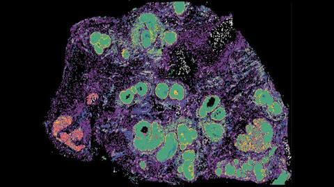
To investigate these interactions, the researchers used single-cell and spatial profiling technologies to examine more than 850,000 cells from normal breast tissue, DCIS and invasive breast cancer samples.
The analysis revealed extensive changes in both cancer cells and their surrounding microenvironment as the disease progressed. It also suggested that some biological features associated with invasive breast cancer begin to emerge earlier than previously recognised.
Uncovering structures linked to tumour growth
Among the findings was the identification of previously unknown structures described as ’cancer stromal loops.’ These ring-like structures, made up of supportive tissue and vascular cells, surround DCIS lesions and appear to create conditions that support tumour growth.
The discovery provides further evidence that tissue surrounding abnormal breast cells may contribute to disease progression, rather than cancer development being driven solely by changes within the tumour cells themselves.
Understanding these interactions could help researchers identify biological features that distinguish lesions likely to become invasive from those that remain non-invasive.
Towards better risk prediction
The researchers believe the atlas could eventually support the development of diagnostic tools to improve risk assessment and help guide treatment decisions for patients with DCIS.
However, further research is needed to validate the findings and establish whether the cellular features identified can reliably predict which lesions will progress.
The team will continue investigating these features to assess their potential use in developing more targeted prevention and treatment strategies, with the aim of identifying patients who need intervention while avoiding unnecessary treatment for those at lower risk.






No comments yet