Image-based, quantitative approaches to study SARS-CoV-2 cell entry and infection: looking for the Achilles’ heel of virus infection
31 March 2022
Shares
- Like
- Digg
- Del
- Tumblr
- VKontakte
- Buffer
- Love This
- Odnoklassniki
- Meneame
- Blogger
- Amazon
- Yahoo Mail
- Gmail
- AOL
- Newsvine
- HackerNews
- Evernote
- MySpace
- Mail.ru
- Viadeo
- Line
- Comments
- Yummly
- SMS
- Viber
- Telegram
- Subscribe
- Skype
- Facebook Messenger
- Kakao
- LiveJournal
- Yammer
- Edgar
- Fintel
- Mix
- Instapaper
- Copy Link
Watch our free on-demand webinar to discover how fully automated imaging and image analysis can provide the unprecedented possibility to follow single viral particles, including for SARS-CoV-2.
About this webinar
SARS-CoV-2 is the causative agent of the COVID-19 pandemic. Understanding the molecular mechanisms of how this virus infects our cells is key to the development of antiviral therapies.
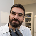
In this webinar, expert speaker Dr Giuseppe Balistreri, Principal Investigator and Adjunct Associate Professor at the University of Queensland, illustrates how to use powerful three-dimensional (3D), automated and quantitative imaging approaches to study how SARS-CoV-2 enters cells of the respiratory tract and neurons. By readapting techniques originally conceived for large screening approaches, Balistreri explains how high-content microscopy can be used for single cell experiments. He covers how fully automated imaging and image analysis using the Yokogawa CellVoyager™ CQ1 provides the unprecedented possibility to follow single viral particles and cellular proteins in millions of cells.
With reagents readily available, software that is easy to use, compact instrumentations and simple protocols, research can be accelerated. These methods also reduce costs, produce unbiased quantitative results and allow any researcher to address a greater number of questions.
Learning outcomes of this webinar
- Understand how to study SARS-CoV-2 cell entry and infection using image-based approaches
- Learn how to readapt high-content microscopes to ‘every-day’ experiments
- Hear about quantitative 3D imaging in an automated fashion
- Explore how automated quantitative imaging can lead to antiviral drug discovery.
Our speaker


Dr Giuseppe Balistreri obtained a master’s degree in molecular biology and microbiology from the University of Palermo, Italy. After moving to Finland, he obtained a PhD in Molecular Virology from the University of Helsinki, focusing on virus replication and virus-induced host membrane rearrangements, in the team led by Professor Leevi Kääriäinen. His research on viruses, particularly virus entry, continued in the research group of Professor Ari Helenius at the ETH Institute in Zurich, Switzerland, where Dr Balistreri developed novel image-based screening approaches working on multiple human viruses. After a short excursion into oncogenic herpesviruses, Dr Balistreri became a principal investigator at the University of Helsinki where he currently works, focusing on the mechanism of SARS-CoV-2 cell entry and spreading and on the development of novel antiviral therapies and improved vaccines. Since 2020, Dr Balistreri is also an adjunct Assistant Professor at the University of Queensland in Brisbane, Australia where he studies how viruses infect and spread in neurons.


