Application note: Tools and protocols for high-content imaging and analysis
Posted: 14 May 2019 | Thermo Fisher Scientific | No comments yet
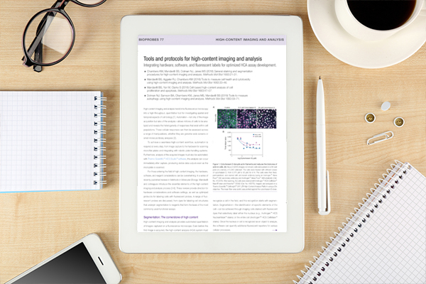
High-content imaging and analysis transforms fluorescence microscopy into a high-throughput, quantitative tool for investigating spatial and temporal aspects of cell biology. Automation – not only of the image acquisition but also of the analysis – allows millions of cells to be analysed and reveals the heterogeneity of responses that exist within cell populations. These cellular responses can then be assessed across a range of manipulations, whether they are genome-wide screens or small-molecule library analyses.
6.high-content-imaging-analysis-software-fluorescent-labels-bioprobes-77-article-tracked-linksRelated content from this organisation
- Application note: Evaluation of hepatic function in 3D culture
- Application note: Hypoxia measurements in live and fixed cells
- Application note: High-throughput imaging and analysis of spheroids
- Application note: Oxidative Stress Measurements Made Easy
- Application note: Understanding Cell Death by High Content Analysis
Related topics
Drug Discovery, Screening, Stem Cells, Target Molecule
Related organisations
High Content Analysis from ThermoFisher Scientific


