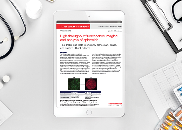
Application note: High-throughput imaging and analysis of spheroids
Posted: 25 September 2019 | Cellular Imaging systems from Thermo Fisher Scientific, High Content Analysis from Thermo Fisher Scientific | No comments yet
High-throughput fluorescence imaging and analysis of spheroids – tips, tricks, and tools to efficiently grow, stain, image, and analyze 3D cell cultures
With optimal cell culture reagents, robust fluorescent reagents and assays, and high-performance fluorescence imaging and detection systems, switching from 2D to 3D cell culture can be easily accomplished even in standard laboratory settings. Because 3D spheroids have a cellular environment and other features that more closely resemble tumors and in vivo models, research on these cultures can provide more relevant data and findings that apply to intact biological systems, enhancing research in drug discovery, cancer biology, and other critical areas.
Related content from this organisation
- Application note: Evaluation of hepatic function in 3D culture
- Application note: Hypoxia measurements in live and fixed cells
- Application note: High-throughput imaging and analysis of spheroids
- Application note: Oxidative Stress Measurements Made Easy
- Application note: Understanding Cell Death by High Content Analysis
Related topics
Assays, Drug Discovery Processes, Genomics, Screening, Stem Cells, Targets, Translational Science


